

AI for Blood Vessels: Russia Develops Digital Assistant to Analyze Arteries
Scientists at Southern Federal University are developing an AI assistant that will help physicians assess the condition of arteries during coronary angiography. The system is designed to reduce the workload on medical professionals and make stenosis assessments more accurate.
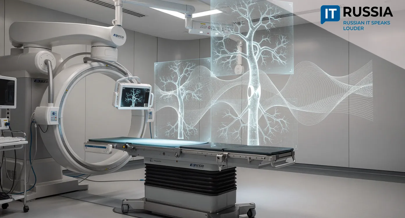
Cardiovascular disease remains one of the most common groups of conditions. One problem faced by cardiologists and endovascular surgeons is stenosis, a pathological narrowing of an artery. It restricts blood flow through the vessel, which can reduce the amount of oxygen reaching organs and tissues. Vascular stenosis can lead to stroke, ischemic heart disease and myocardial infarction.
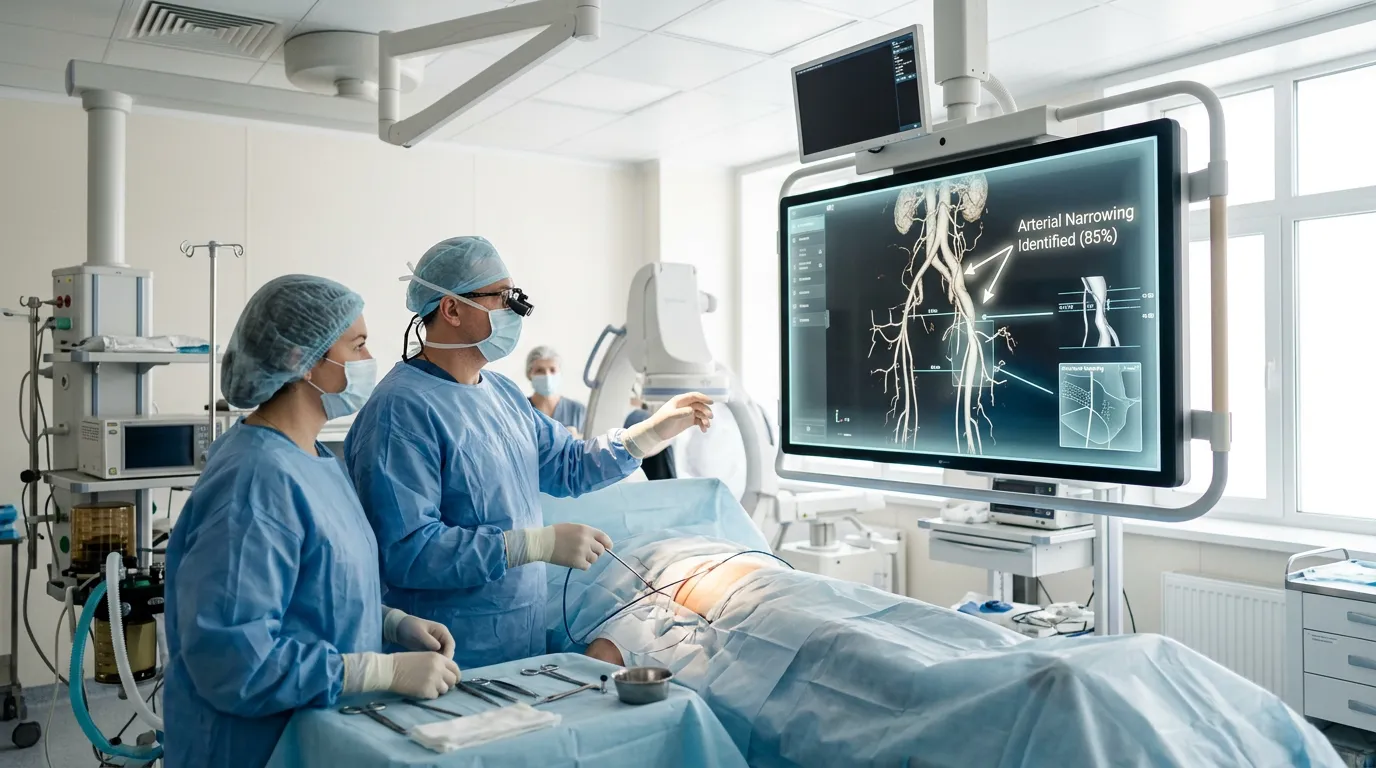
To assess the condition of the coronary arteries, physicians use invasive coronary angiography. During the procedure, a contrast agent is injected into the vessel, and X-ray equipment captures images of blood flow. The physician analyzes those images to determine where a narrowing is located, how severe it is and how the vessel’s geometry changes. But this is a complex process because it requires reviewing numerous images captured at different points in time and from different projections. Scientists at Southern Federal University have found a way to delegate this work to AI.
A New Way to Analyze Vascular Images
The “Intelligent Assistant for Endovascular Surgeons” project was one of the winners of Southern Federal University’s competitive selection of artificial intelligence projects. It is being developed at the request of Taganrog City Clinical Emergency Hospital No. 5.
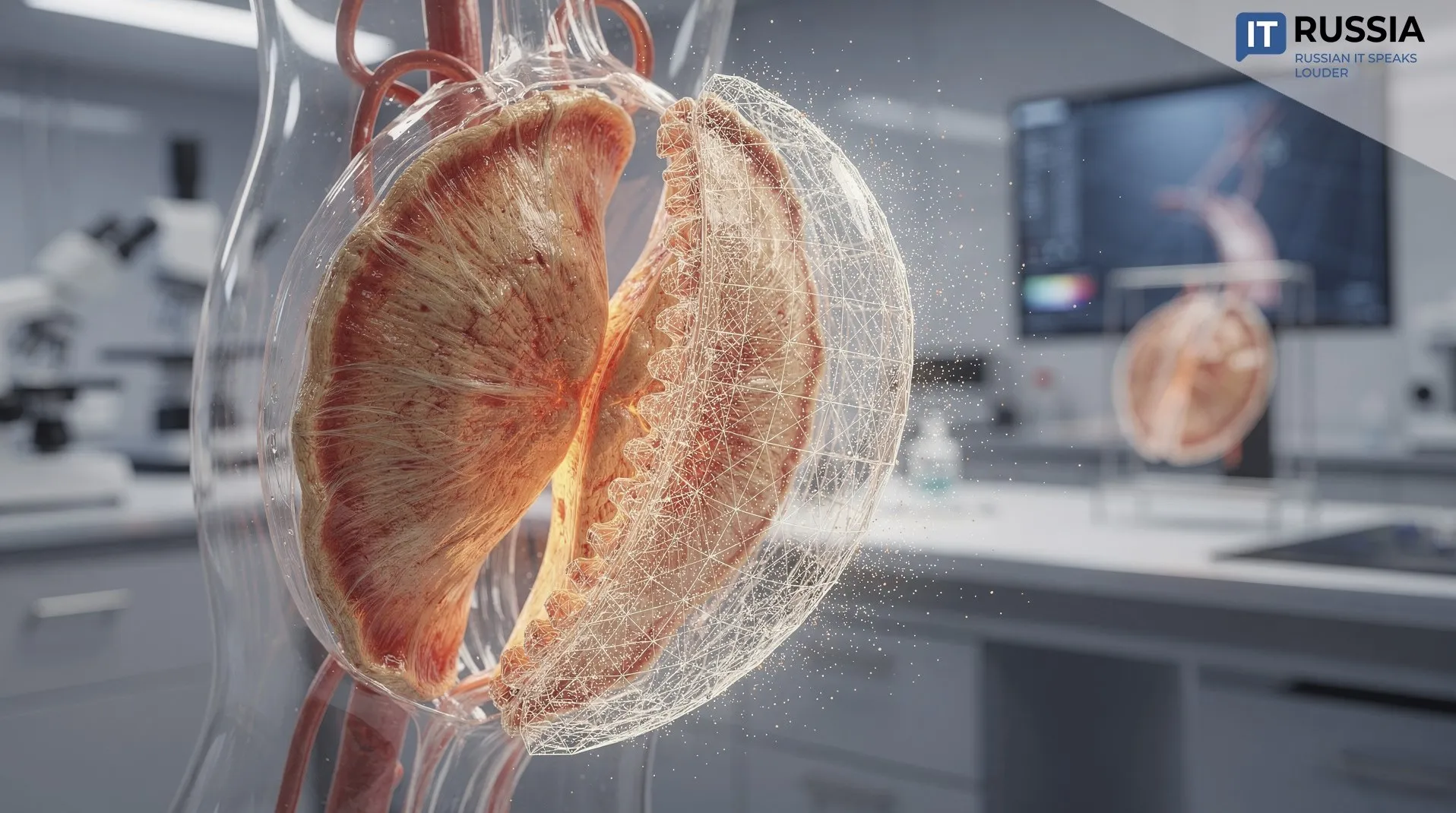
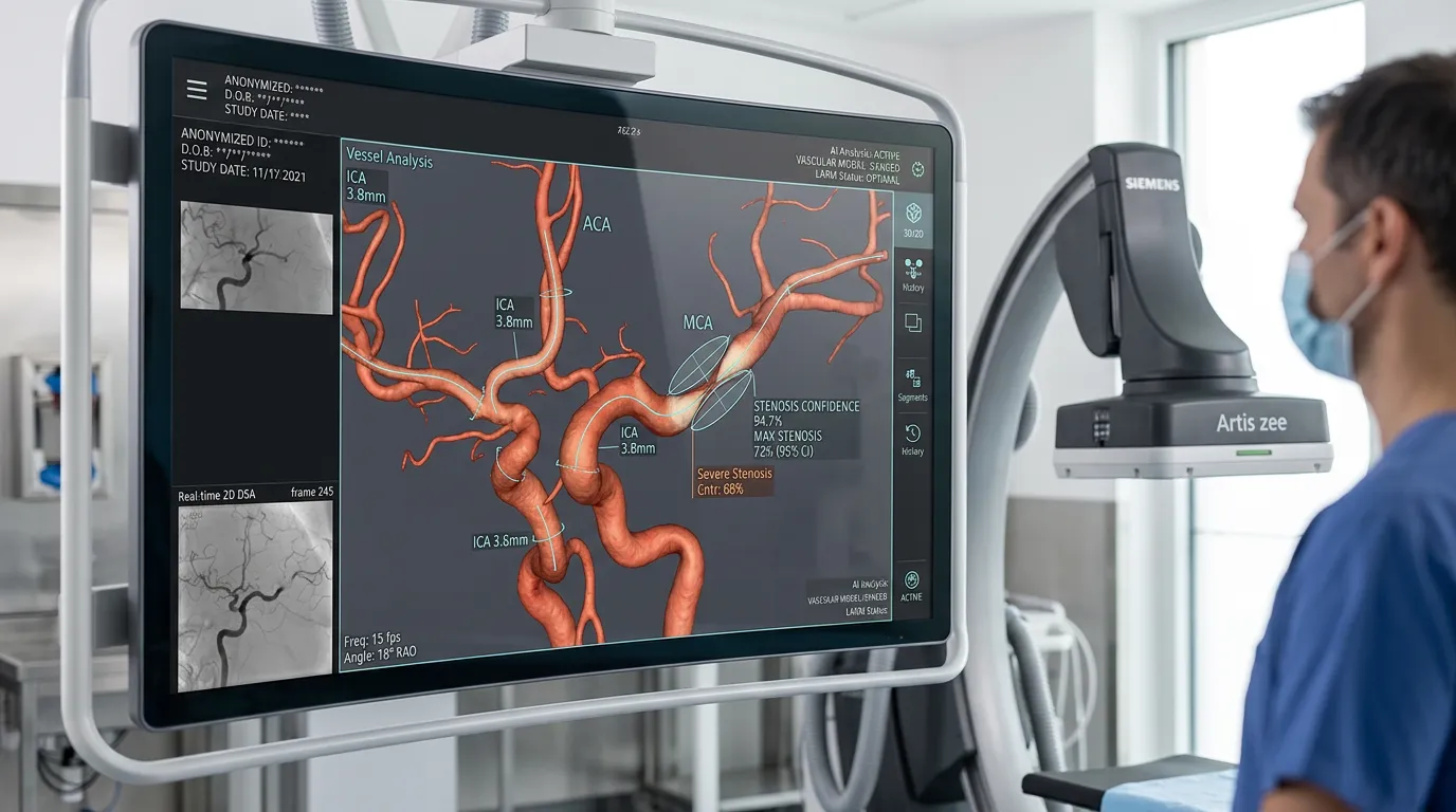
The key feature of the system is that it is designed to analyze angiography as a single video sequence. A 3D model can be used to assess the geometry of a vascular segment, pinpoint the location of narrowings, match images captured from different projections and prepare data for quantitative stenosis assessment. This fundamentally changes how the data can be processed. A computer can track how the vessel changes over time, link its contours and centerlines, and then reconstruct the vascular network and its 3D model. In other words, the AI builds a complete picture of the structure of a specific section of the vasculature and whether it carries a risk of narrowing.
Why a 3D Model Matters
As Dmitry Safonov, acting chief physician of City Emergency Hospital No. 5, explained, cardiovascular diseases remain widespread, and the number of patients with such diagnoses continues to rise each year. This puts additional pressure on medical professionals, which can reduce diagnostic accuracy, increase the risk of medical errors and contribute to burnout among healthcare workers.
The assistant could reduce the workload on physicians, improve the reproducibility of results and reduce subjectivity in assessments. For patients, that could mean more accurate diagnoses and fewer diagnostic errors.
Students Put to Work
The project is also being used in Southern Federal University’s educational programs. A total of 250 students participated in data annotation, most of them enrolled in TOP-DS and DS programs. They added structured labels to the original images and videos. Before that, leading experts in coronary angiography conducted a series of training sessions for faculty members and senior students serving as mentors.
As Alexander Kozlovsky, a senior lecturer in the Department of Computer Engineering at Southern Federal University’s Institute of Computer Technologies and Information Security, explained, the students gained hands-on experience solving practical problems. Those who performed best were invited to join the project’s core team.
Developing Medical AI
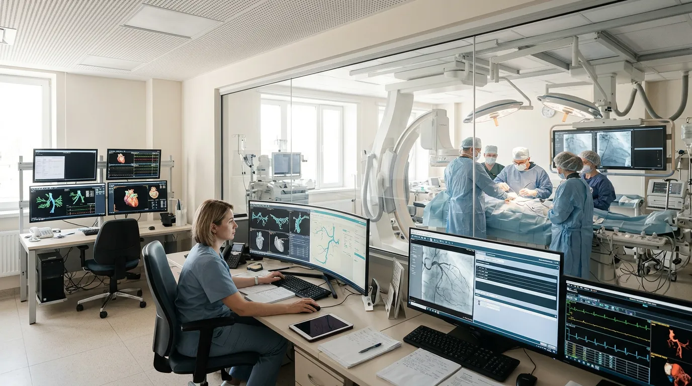
Russian medical AI systems can already handle a wide range of tasks, such as detecting signs of disease in X-ray and tomographic images. The “Intelligent Assistant for Endovascular Surgeons” project represents a different stage in that development. Here, the algorithm will have to work with several types of information at once, account for the sequence of images, reconstruct the three-dimensional structure of blood vessels and produce quantitative measurements.
In the future, systems like this could be used for more than initial analysis. They could be developed to support planning of endovascular procedures, compare studies over time and monitor treatment outcomes. For Russia’s IT industry, similar projects also matter because they enable the development of specialized technologies based on real-world needs identified by hospitals.