

Penza Researchers Develop Simulator for Detecting Defects in X-Ray Images
An interactive web-based simulator allows medical students and resident physicians to learn how to identify defects in X-ray images.
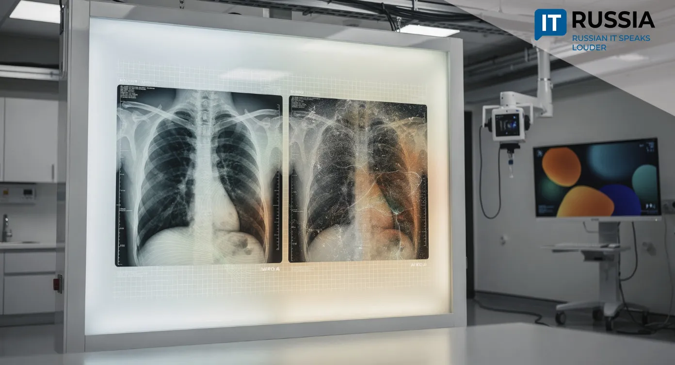
The worse the image quality, the harder it is to spot pathology in time. Equipment condition, exposure settings, radiation dose, quantum noise – all of these factors matter. That means a medical professional must not only recognize disease, but also understand which parts of an image reflect the actual clinical picture and which result from technical distortion.
Researchers at Penza State University (PSU) have proposed their own approach. Their new simulator helps users distinguish technical artifacts from changes in the body.
Matching the Reference
The system has three components. The first is a database that stores X-ray images, quality metrics and related information. The next is a metrics-calculation subsystem. It automatically evaluates images using multiple quantitative measures, including the signal-to-noise ratio, image gradients, and the difference between maximum and minimum brightness. All calculations are based on pixel data and use rigorous mathematical formulas. This makes the results objective and reproducible.
The final component is the training and testing subsystem. It generates exercises using defective versions of reference images. A radiologist uploads images to the database, and an expert selects the best ones and marks them as references. The system then creates distorted copies that simulate real-world defects. Metrics are calculated for each copy as well.
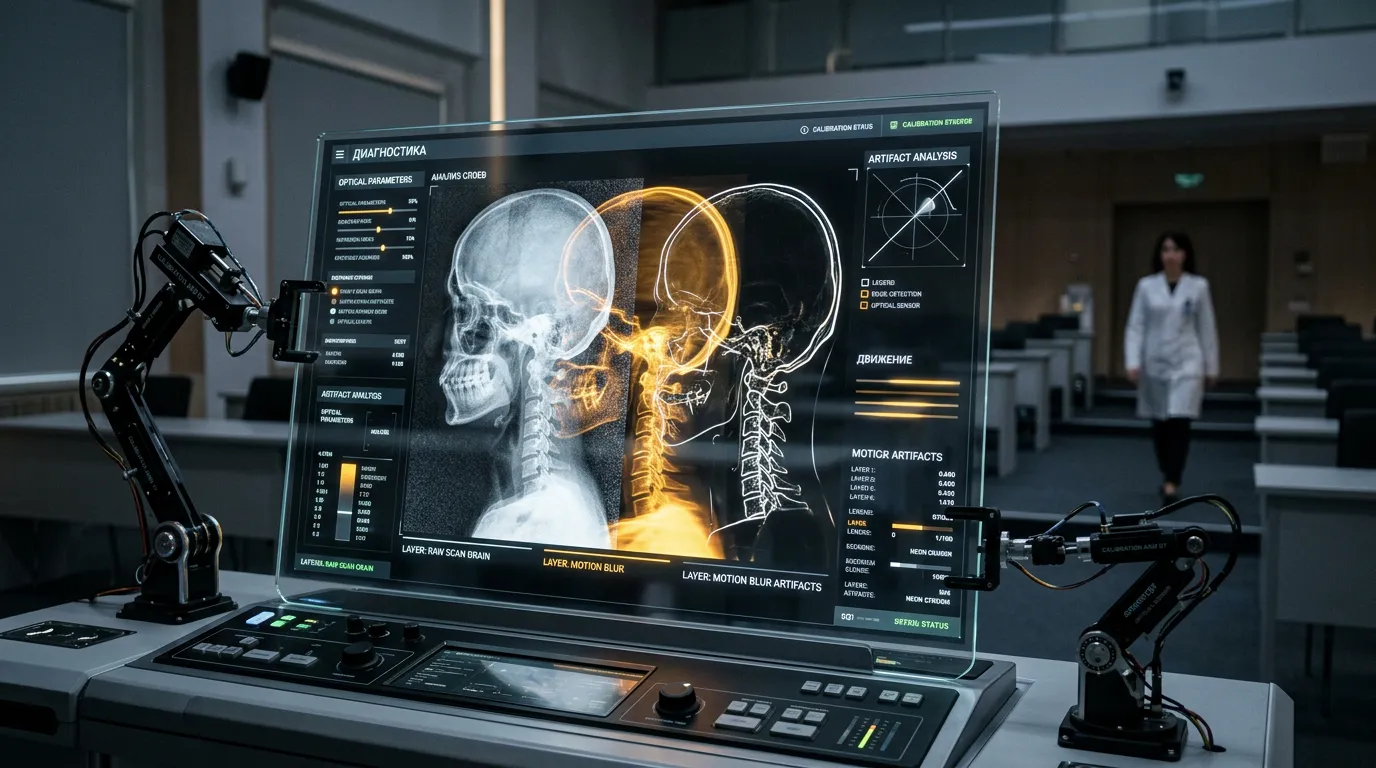
200 Cases – 100% Accuracy
All students need is a browser and internet access to train with the simulator. They receive exercises in which they see the reference and distorted images simultaneously. The system provides immediate feedback, helping students connect visual features with numerical measurements. All responses are stored in the database, where both instructors and students can work with them.
At the PSU Medical Institute, researchers used the simulator to evaluate 200 test cases. Starting with five reference images covering the chest, extremities and abdominal cavity, the system generated 10 distorted copies for each of four types of defects. With carefully selected metric thresholds, it correctly classified the type of defect in every case.
Millions of Images
AI technologies for medical image analysis have been developing actively in Russia for several years. In 2022, clinical decision-support systems were developed that automatically search for signs of pathology in X-ray and CT scans.
Russian regions began adopting image-analysis technologies in 2023. In the Arkhangelsk Region, for example, AI systems were used to process diagnostic imaging studies. During 2024, AI was used to analyze more than 300,000 X-ray images of residents in the region.
By 2025, reports were already citing millions of medical imaging studies in Moscow processed with AI algorithms. In January 2025 alone, 245,000 X-ray and fluorography examinations were performed with clinical decision-support systems across 20 Russian regions. In 10 regions, AI was used for more than 15,000 chest CT examinations.
“There has been a sustained trend toward greater use of clinical decision-support systems. In the first month of this year alone, the volume of outpatient mammography studies using artificial intelligence reached nearly 90,000 examinations, 37% higher than in the same period last year,” Olga Tsareva, deputy chair of Russia’s Federal Compulsory Medical Insurance Fund, said last year.
Since 2025, additional funding has been allocated for the use of clinical decision-support systems in X-ray imaging, fluorography and chest CT. PSU’s development extends this work, but with a focus on training medical professionals.
Future Scenarios
Medical AI will likely expand its training scenarios next, covering CT, MRI and mammography, along with personalized training programs and integration with medical universities’ educational platforms. At the same time, simulators could be combined with clinical decision-support systems. PSU researchers have already introduced an intelligent chest X-ray analysis system called Radex. It evaluates chest X-rays and detects signs of 18 pathologies.
In the coming years, comprehensive educational platforms could also emerge, bringing simulators, AI and real-world medical scenarios together. For now, students at the PSU Medical Institute are closely examining X-ray images provided by their simulator assistant.