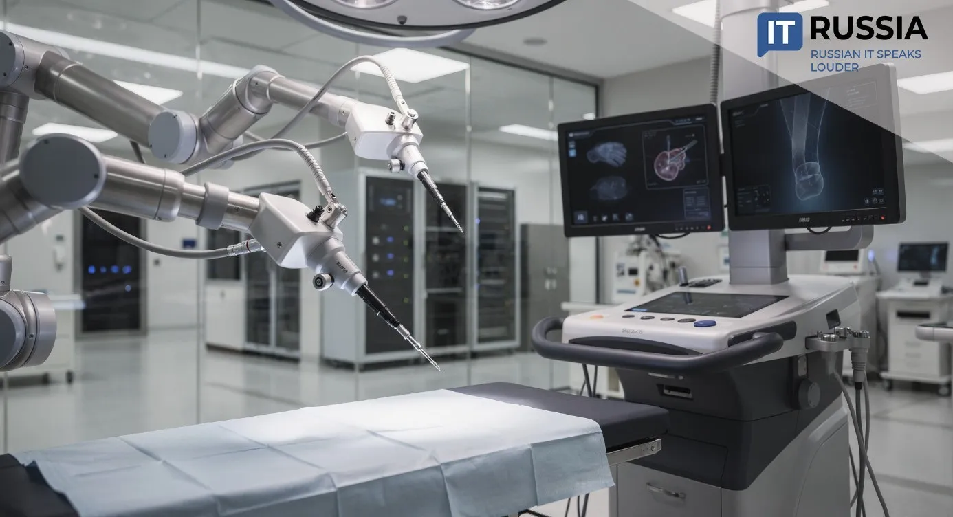
Precision as Standard: Russian Algorithm Boosts CT Imaging Resolution
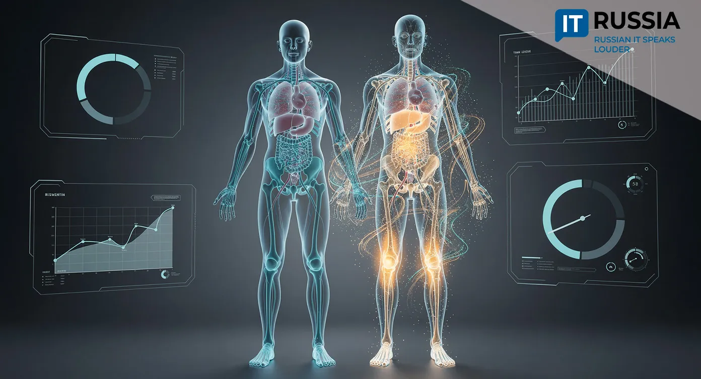
Russian researchers have developed a cost‑efficient method for modernizing medical imaging systems. The software‑driven approach enables older CT systems to deliver ultra‑sharp diagnostic images through dual‑scan reconstruction – improving diagnostic accuracy at scale without requiring major capital upgrades.
Mathematics From the Far East
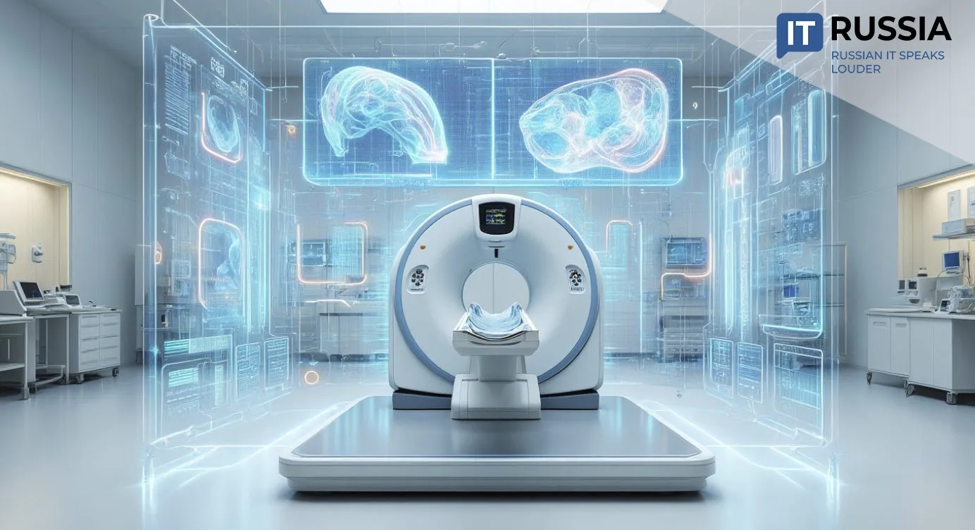
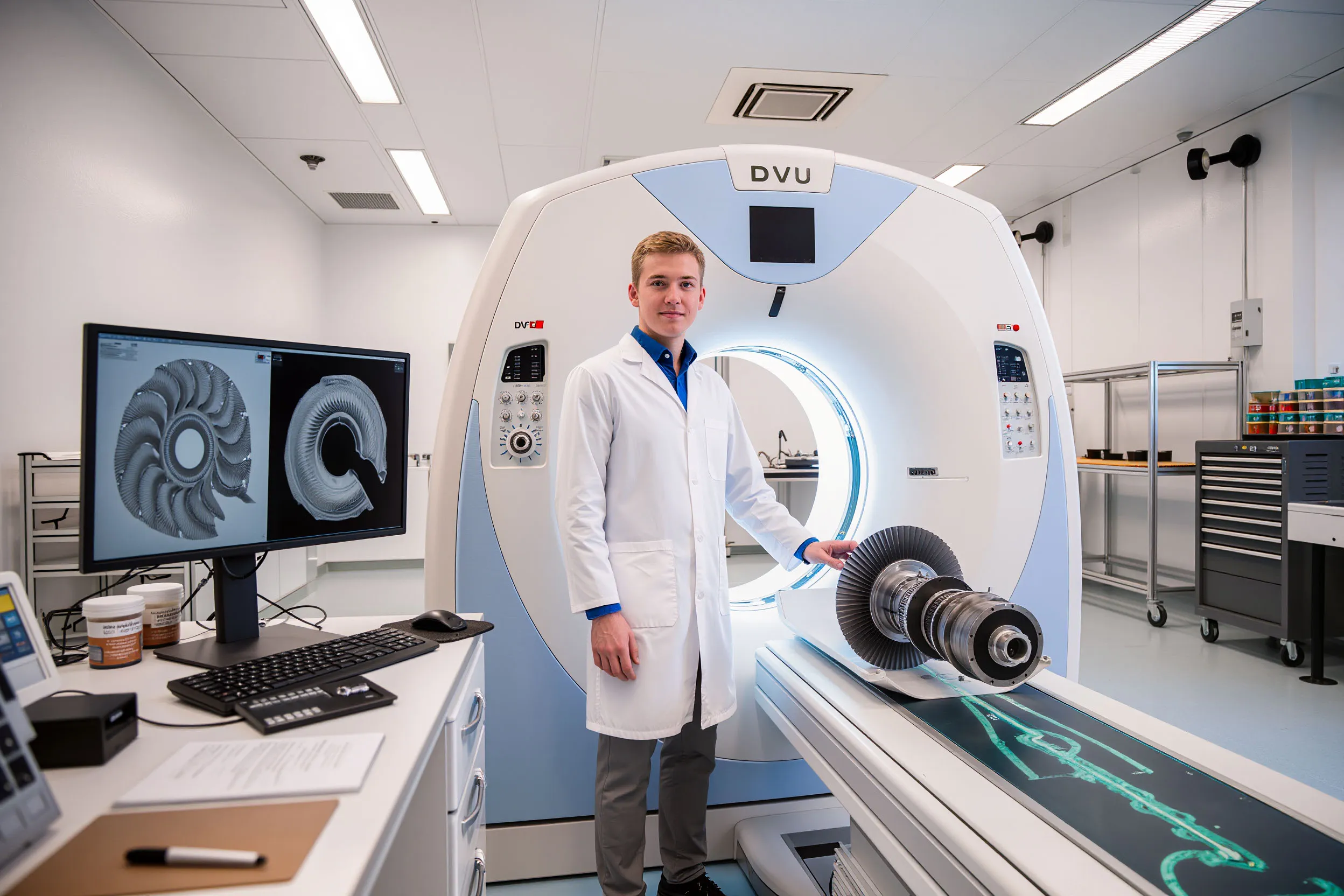
Improving diagnostic precision does not always require purchasing new hardware. A mathematical method developed in Russia’s Far East significantly enhances CT system performance through a software retrofit. A joint research team from Far Eastern Federal University and the Institute of Applied Mathematics of the Far Eastern Branch of the Russian Academy of Sciences detailed the new mathematical algorithm in a series of scientific publications.
Its foundation is paired imaging with minimal variation in acquisition parameters – for example, slight changes in X‑ray beam width. When two mildly blurred scans of the same object are captured, a dual‑scan reconstruction algorithm mathematically extrapolates and subtracts imaging noise, producing a final image with far greater detail than conventional single‑scan imaging. The CT hardware remains unchanged – only the data‑processing logic evolves.
Significance for Russia
In a global market historically dominated by foreign manufacturers of high‑tech medical and industrial imaging systems, competing in hardware alone demands enormous time and resources. Russia has adopted a more pragmatic approach: rather than building new scanners from scratch, it aims to dramatically increase the informational output of existing systems – both those already in domestic clinics and those that could be retrofitted abroad.
The project demonstrates how decades of fundamental mathematical research conducted in Russian Academy of Sciences institutions translate into practical clinical and industrial applications. This strengthens Russia’s profile as a center of intellectual export, where algorithms and ideas become high‑value assets.

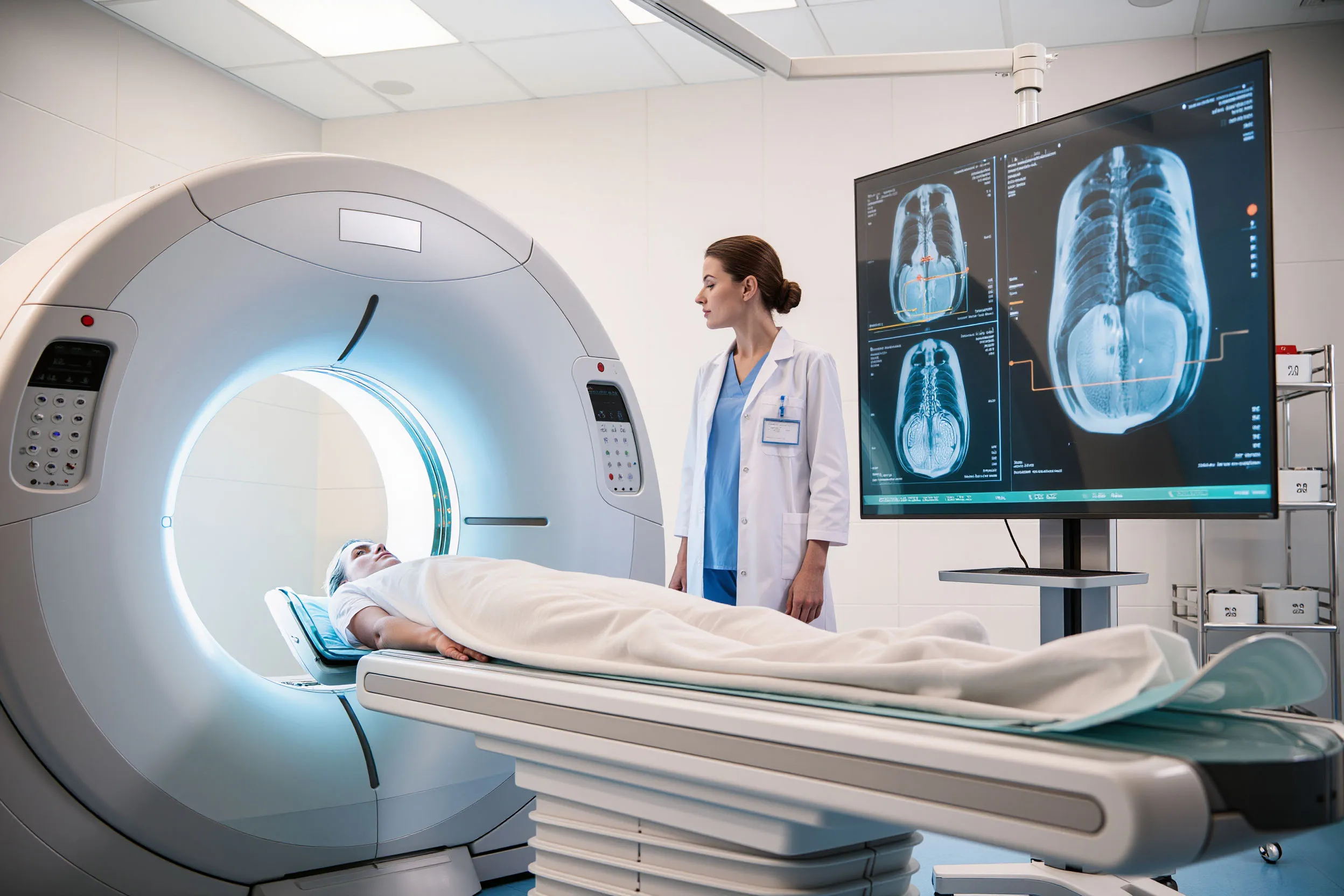
Deploying such software across Russian clinics and diagnostic centers could be both large‑scale and relatively fast. Even older‑generation CT systems would be able to produce images comparable in clarity to new‑generation models. For national healthcare, this offers a path to significantly raising diagnostic standards without the massive budget shock of replacing entire equipment fleets.
A Stimulus for the IT Sector
The method forms the basis for specialized software that Russian IT companies can further develop and commercialize. A new niche is emerging at the intersection of medicine, industrial imaging, and software engineering – a segment where domestic developers may hold strong competitive advantages.
Its export potential may exceed domestic demand. Hundreds of thousands of CT systems operate worldwide. A software licensing model that allows hospitals or industrial sites to improve image quality without spending millions of dollars on new hardware is highly attractive.
For developing countries, the approach offers a practical way to modernize diagnostic infrastructure using existing equipment. For major clinics and corporations, it helps extract maximum value from current assets and improve study precision. For CT manufacturers, it presents a potential technology for licensing and integration as a competitive differentiator.
From Pixels to Predictions
The implications extend well beyond producing clearer images. The technology reshapes clinical workflow. Higher resolution enables physicians to distinguish smaller structures with lower contrast differences. In oncology, this may allow visualization of earlier or smaller tumor sites or subtle tissue changes. In neurology, it can improve visibility of fine vessels or early demyelination. In practice, it shifts diagnostics toward earlier – and therefore more treatable – stages of disease.

Greater detail can also reduce reliance on invasive diagnostic procedures, such as exploratory surgery or painful biopsies, when non‑invasive imaging provides sufficiently comprehensive information.
In surgery and radiotherapy, precise tumor contours and clarity regarding small vessels and nerves are essential for minimizing complications. An algorithm that increases image detail directly supports better treatment outcomes.
High‑quality digital imaging also provides a foundation for remote multidisciplinary consultations. Standardized, high‑resolution images facilitate collaboration among clinicians across cities and countries.