Neural Network in Russia Is to Learn to “Read” Jaws From Imaging Scans
The project could eventually change the approach to diagnostics in dentistry.
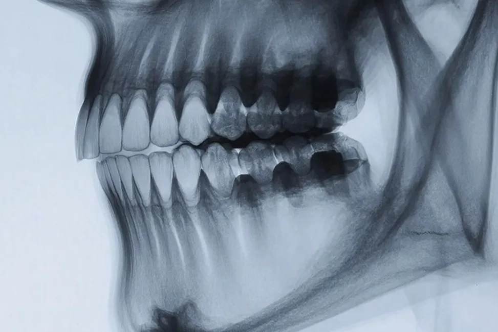
At Yaroslav-the-Wise Novgorod State University, researchers are working on a project that could reshape dental diagnostics. The focus is a service that automatically analyzes orthopantomograms — complex panoramic jaw X-ray images.
The project’s author is Lyubov Skladnik, a master’s student at the university’s Polytechnic Institute. The university’s press service told IT Russia.
A Standard Method, With a Catch
Panoramic X-rays of the maxillofacial area allow dentists to assess the condition of teeth, bone tissue, and jaw joints, as well as detect inflammation, cysts, hidden carious processes, and other pathologies. The method is highly informative and has become a standard tool in modern dentistry. The problem is that orthopantomograms can be accurately interpreted only by highly qualified specialists, and analyzing a single panoramic image can take considerable time.
There is also an element of subjectivity. Even experienced doctors may interpret the same image differently, especially when changes are subtle or at an early stage. As a result, there is always a risk of missing pathology in its early phases.
A Neural Network to ‘Read’ the Image
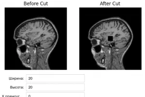
The task is complex: to develop a method for automatically identifying diagnostically significant features in panoramic images. In other words, the system must not only ‘see’ teeth and bones, but analyze their structure, detect pathologies, and correlate them with clinical criteria. This will be handled by a neural network. The project plans to use a pre-trained ResNet model, which will then be fine-tuned on datasets of orthopantomograms from open sources.
Preparing for Clinical Use
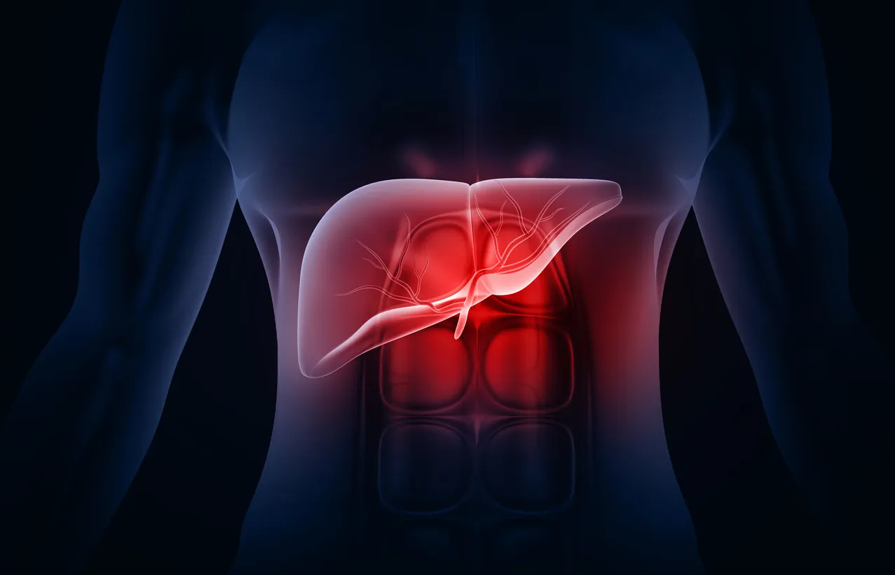
In theory, the technology could also be applied to X-rays of bones and internal organs, as well as to ultrasound imaging. The system will not replace doctors, but is intended to serve as a convenient decision-support tool.
The project was presented at the international scientific and practical conference Digital Horizons: Modern Challenges of Computer Science and Computing Technology, held at the university. Testing is planned at a clinic in Veliky Novgorod. If successful, the system will be introduced into clinical practice.
Earlier, we reported that Saint Petersburg Electrotechnical University presented an innovative dental solution — a compact device for rapid early-stage caries diagnosis. Unlike traditional X-ray systems, the new device uses safe semiconductor lasers, allowing examinations to be performed directly in the dentist’s chair without harm to patients.