Russia has developed an app that can assess the quality of X-ray images
Russian researchers have developed a cross‑platform app that evaluates the quality of X‑ray images, promising faster diagnosis, lower radiation exposure, and reduced clinical costs

Medicine and Digital Health
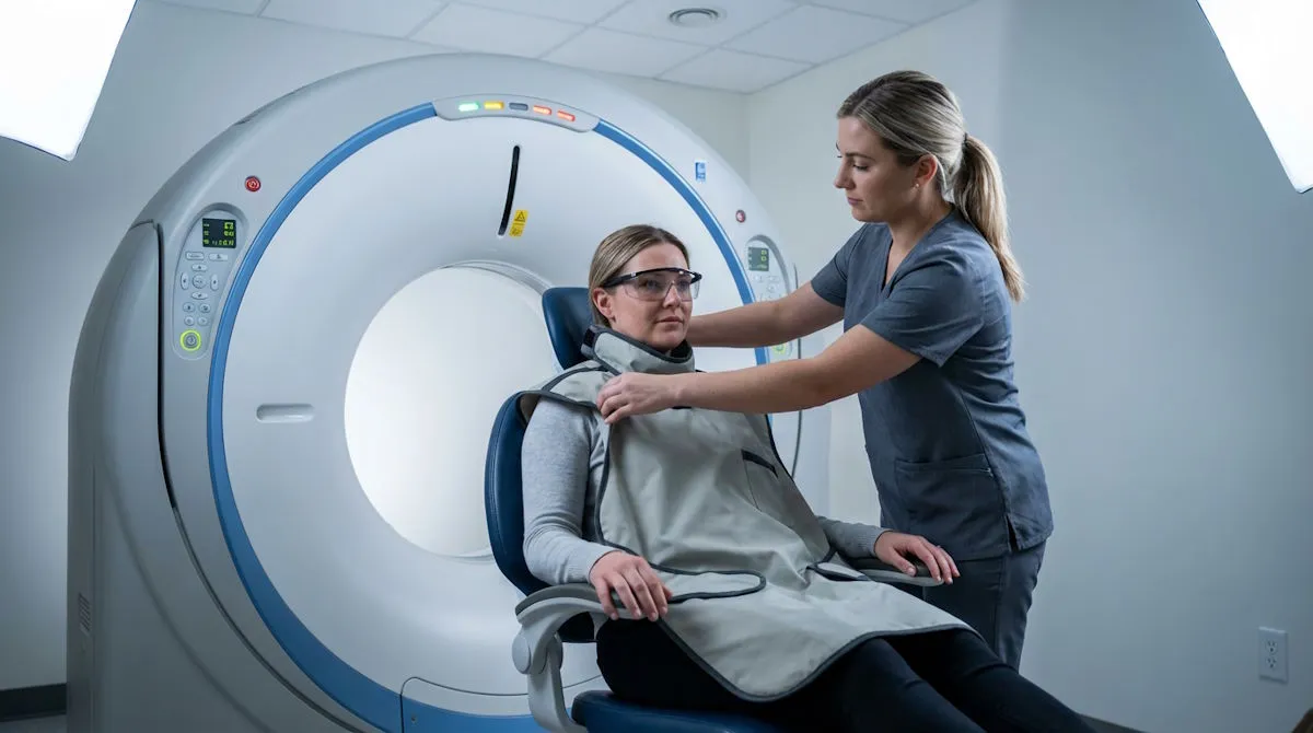
A research team from Penza State University has created a cross‑platform application capable of automatically assessing the quality of X‑ray images, according to the Russian Ministry of Science and Higher Education. The system is designed to reduce the number of repeated radiological examinations — a problem that affects between 5% and 15% of all X‑rays, often due to insufficient image quality.
The app, developed by Professor Leonid Krivonogov from the Department of Medical Cybernetics and Informatics and medical student Darina Ulybina, accelerates diagnosis and helps cut procedural costs. The tool is intended not only for radiologic technicians and radiologists but also for use in medical schools.
An AI‑Driven Image‑Quality Assistant
The platform incorporates a suite of mathematical algorithms and includes 11 metrics for evaluating imaging quality.
Each metric has its own scoring system that compares uploaded X‑rays to a reference standard. Physicians can also set custom evaluation parameters.
For each clinical case, the app selects the most appropriate preset, producing clearer, more anatomically detailed images through individualized processing.
Quality Control Powered by Data
After analyzing about 100 X‑ray images from open data repositories, the developers found that the app accurately identifies distorted images and flags incorrect preset selections. The platform can also generate a full report on image‑quality assessment — a useful feature for clinical workflow and radiology education.