Minute-Long Diagnosis: Novosibirsk Endoscope With AI to Detect Cancer Without Pain or Biopsy
Russia is developing a new method for diagnosing gastrointestinal diseases. Scientists in Novosibirsk are creating a laser microscope that can be built into an endoscope. The device will make it possible to detect cancer at an early stage, while artificial intelligence analyzes the resulting images.
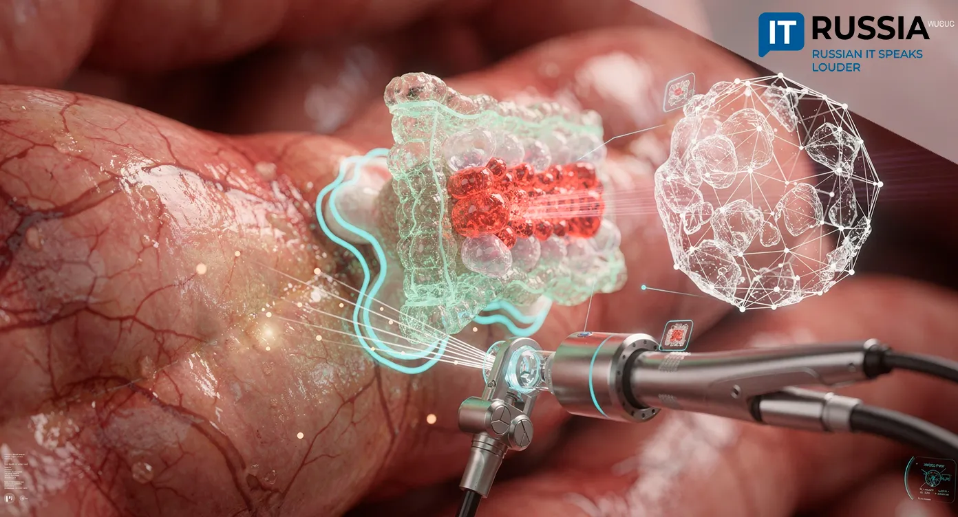
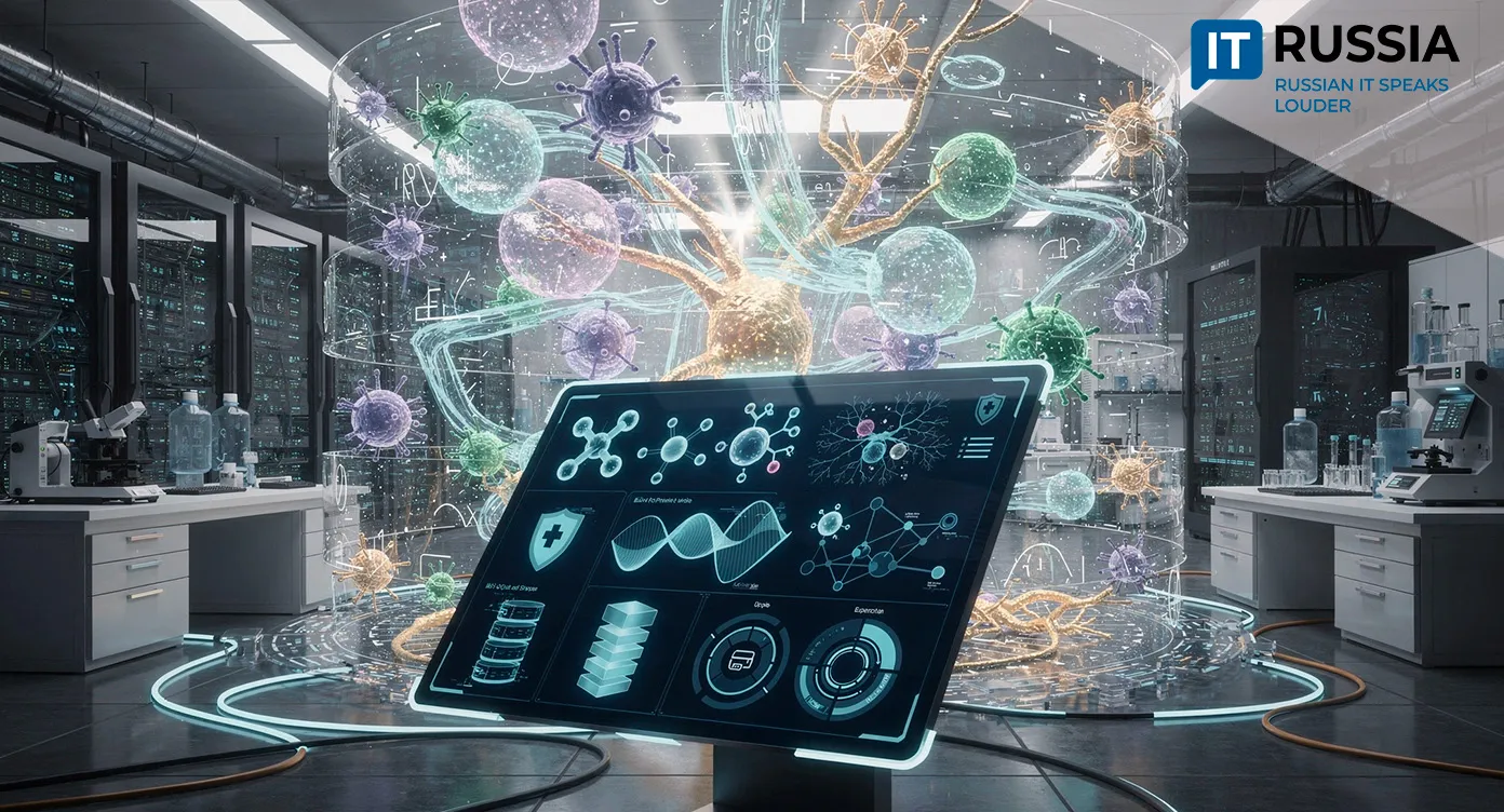
Researchers from Novosibirsk State University, the Institute of Automation and Electrometry of the Siberian Branch of the Russian Academy of Sciences and the Institute of Molecular and Cellular Biology of the Siberian Branch of the Russian Academy of Sciences are working on a new diagnostic method for gastrointestinal diseases. The technology is built around a portable multiphoton microscope integrated into a standard endoscope.
Today, when doctors need to determine whether a patient has stomach or intestinal cancer, they usually have to perform a biopsy. The procedure is far from comfortable. A physician removes a piece of tissue and sends it to a laboratory, with results arriving days later. The process is painful, unpleasant and time-consuming. If several suspicious areas are found, the procedure may have to be repeated multiple times.
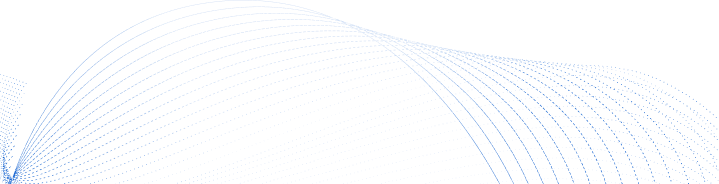
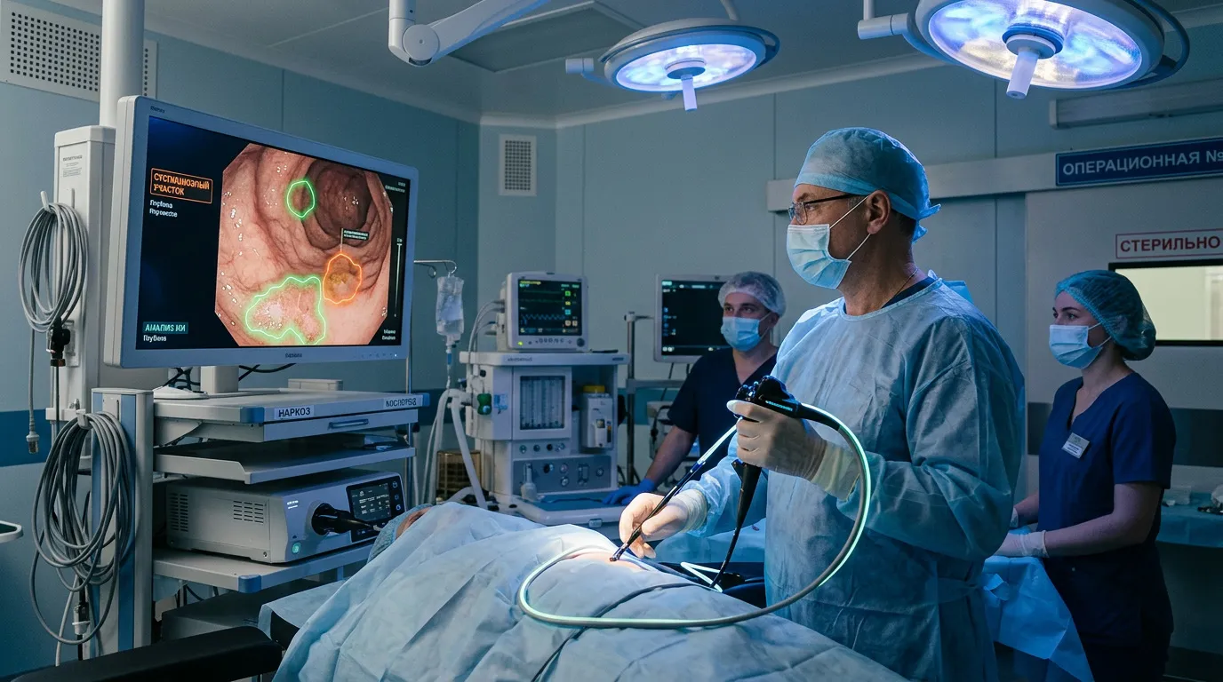
The Novosibirsk method works differently. A doctor inserts an endoscope equipped with a miniature laser microscope, illuminates tissue with infrared radiation and immediately receives a three-dimensional cellular-level image in real time.
How the Technology Works
Conventional microscopes rely on visible light, which does not penetrate deeply into tissue. The Novosibirsk team instead uses a femtosecond laser – ultrashort, high-powered infrared pulses. That type of radiation passes through tissue more effectively without burning it. Because fluorescence requires two photons simultaneously, the method can excite the natural fluorescence of cells without damaging the sample.
The resulting images are analyzed by a neural network. Using previously studied examples, researchers are training AI to distinguish healthy tissue from diseased tissue and identify characteristic signs of illness within three-dimensional structures. What once could only be identified by an experienced morphologist examining biopsy samples under a microscope can now be recognized automatically by the algorithm during the procedure itself.
What It Means for Patients
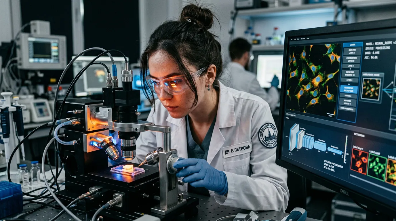
For patients, the main advantage is painless diagnostics. No tissue pinching, no waiting and no anxiety over delayed results. Physicians can see the findings immediately and make decisions on the spot if a problem is detected.
It is well known that early cancer detection directly affects survival rates. For stomach cancer diagnosed at stage one, five-year survival reaches 70% - 90%. At stages three and four, that figure falls to 10% - 30%. The new method could identify precancerous changes long before patients notice symptoms.
The approach could also reduce false alarms. Traditional endoscopy is ultimately based on visual assessment. A physician examines the stomach wall through a camera and decides whether an area appears suspicious. Even highly experienced specialists cannot see what lies beneath the surface. The laser microscope, however, looks inside tissue at the cellular level, while a neural network trained on thousands of examples filters out questionable cases.
What It Means for Russia
For the healthcare system, the technology could lower diagnostic costs while speeding up the process. Biopsies require disposable instruments, laboratory reagents, pathology work and specialized staff. A laser endoscope, by contrast, represents a one-time investment in equipment that can then be reused repeatedly without ongoing consumable costs.
Regions facing shortages of qualified morphologists could gain access to a tool that does not depend on human variability. The neural network performs consistently whether it is used in Novosibirsk or in smaller cities where recruiting highly specialized experts is difficult.
The development is also fully Russian – from physical optics to the AI software itself.
What It Means Globally
Comparable technologies already exist internationally in the form of confocal laser endoscopy. Those systems, however, require intravenous fluorescent dyes that can trigger allergic reactions. The Novosibirsk method does not need additional contrast agents and instead relies on the natural fluorescence of tissue.
Femtosecond lasers are already widely used in ophthalmology for vision correction, but their application in gastrointestinal endoscopy remains an emerging field. Russian scientists are positioning themselves among the leaders by proposing not just a laboratory prototype, but a portable device that can be integrated into a standard endoscope.
For countries without extensive early cancer-screening infrastructure, such a device could make large-scale population screening possible without massive spending on laboratories and specialized personnel.
Future Prospects
The project is currently in the laboratory research stage. Scientists are working with cell cultures, organoids (artificially grown mini-organs) and mice. At the same time, the neural network is being trained on existing examples of healthy and diseased tissue.
The next step is the creation of a working prototype of the portable laser microscope that can be integrated into an endoscope. After that will come clinical trials involving volunteers. The technology could make it possible to catch cancer before metastases develop, when treatment can still avoid aggressive chemotherapy and major surgery.